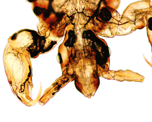
Answer: Strongyloides stercoralis
This appearance of motile larvae on agar culture is classic for strongyloidiasis and fits with the patient's history of immunosuppression and enterococcal meningitis. Meningitis in strongyloidiasis occurs when invasive L3/filariform larvae penetrate the intestinal wall and travel through the blood stream to the CNS, dragging gut bacterial with them. Escherichia coli is the most common bacteria to cause meningitis in this setting, but other Gram negative bacilli and gut flora have also been reported.
It's important to note that Strongyloides agar culture detects ALL motile larvae found in stool, including hookworm larvae that have hatched from their eggs. Therefore, it is important to also examine the morphology of the worms to differentiate Strongyloides larvae from other similar appearing larvae. In particular, Strongyloides L1/rhabditiform larvae have a shorter buccal canal than hookworm L1 larvae, and the L3/filariform larvae have a notched tail, whereas the hookworm L3 larvae have a pointed tail.
Sunday, July 29, 2012
Subscribe to:
Post Comments (Atom)


No comments:
Post a Comment