Here is this week's case:
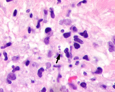
The following is an hematoxylin and eosin stained skin biopsy taken from a mission worker who just returned from the Middle East. He presented with a 5 cm, non-healing ulcer. The biopsy shows numerous small (2-3 micron) objects inside macrophages. The arrows point to a defining feature of the organisms. Identification? Note that the organisms are scant, and hard to make out, even under 1000x oil immersion. What type of slide preparation is better for showing the morphology of this parasite? Based on these images, what is your differential diagnosis? And finally, what special stain would help you exclude some of the other possible diagnoses? (CLICK ON IMAGES TO ENLARGE)


3 comments:
From the history as well as the appearance of the organisms, my vote is for Leishmaniasis.
pathresident
I am a lecturer working in the Department of Parasitology in the Faculty of Medicine, University of Peradeniya, Sri Lanka. We have no way of identifying parasites in tissue sections. Wonder whether you could help us in any way? my email: dhilmaa@yahoo.com
In the description it says they were seen in macrophages, the only kinetoplastid parasite with macrophages replication is leishmania and since this is in humans its the amastigote form of leishmania
Post a Comment